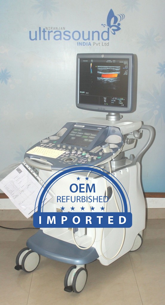
In India we supply/support "The Pre-natal Diagnostic Techniques (PNDT) Act 1994 registered center only
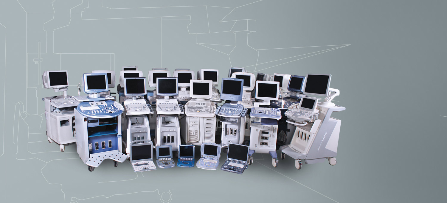
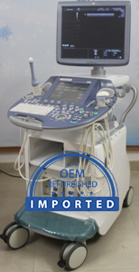
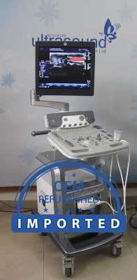
Recent Products
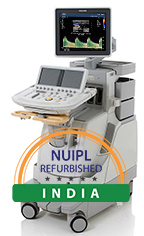
Upcoming Machines
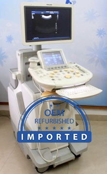
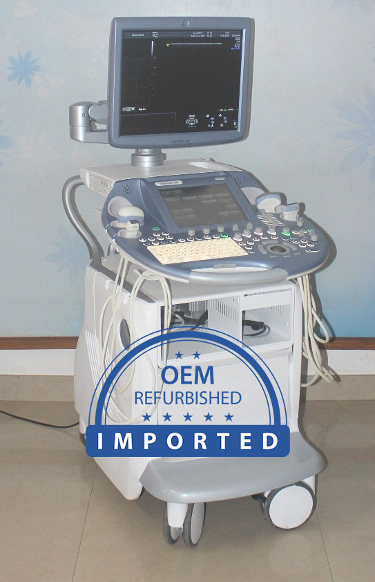
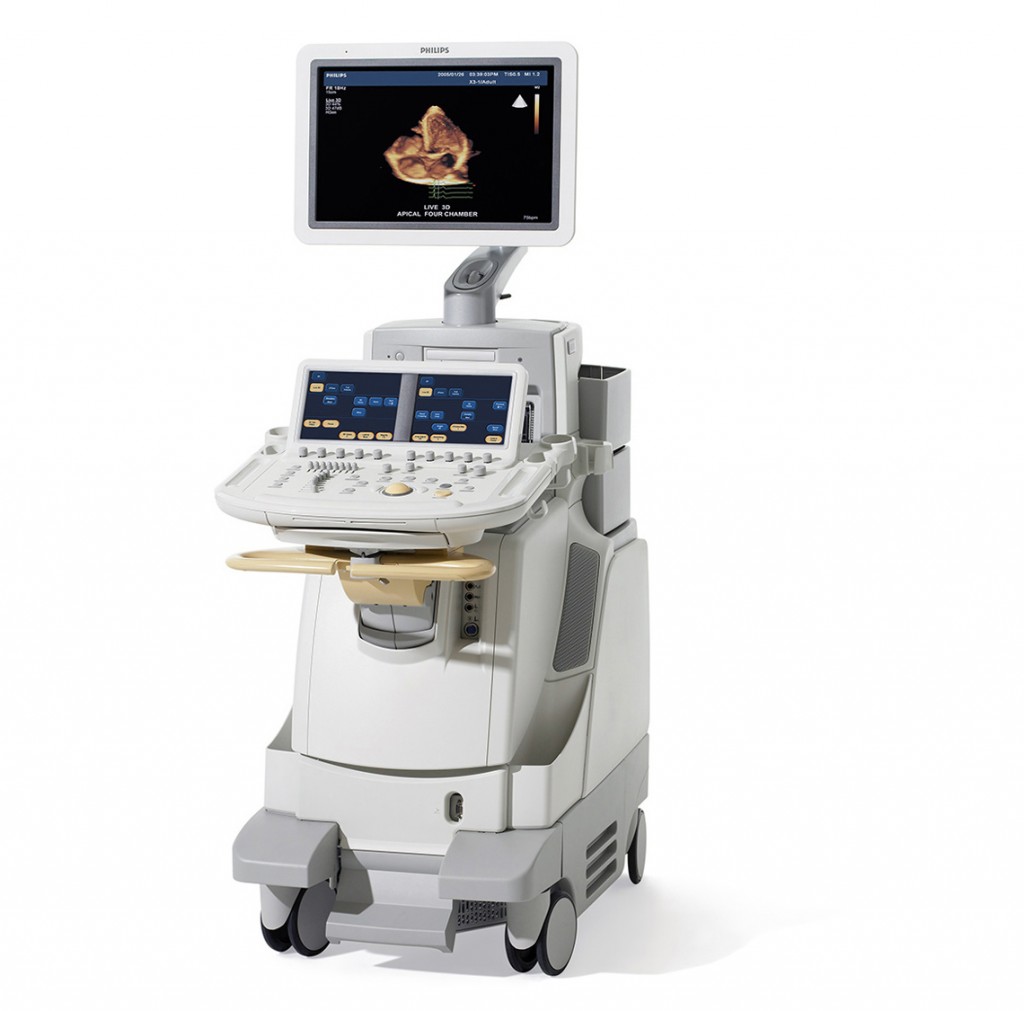
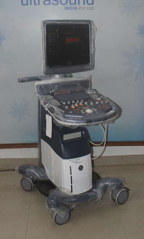
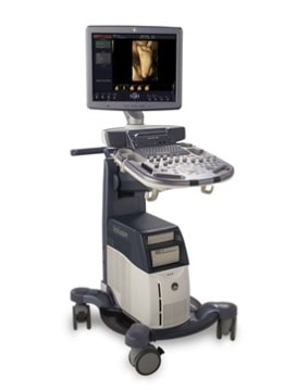
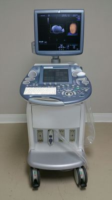
PHILIPS IU22 G Cart
Year of Manufacture : May 2012
Software Version : 6.3.6.
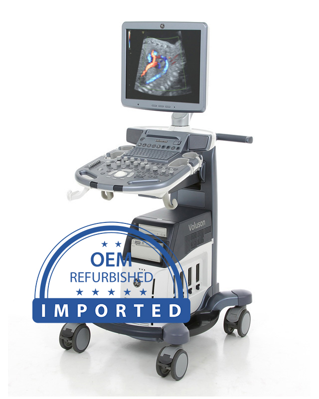
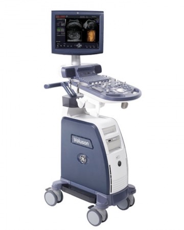
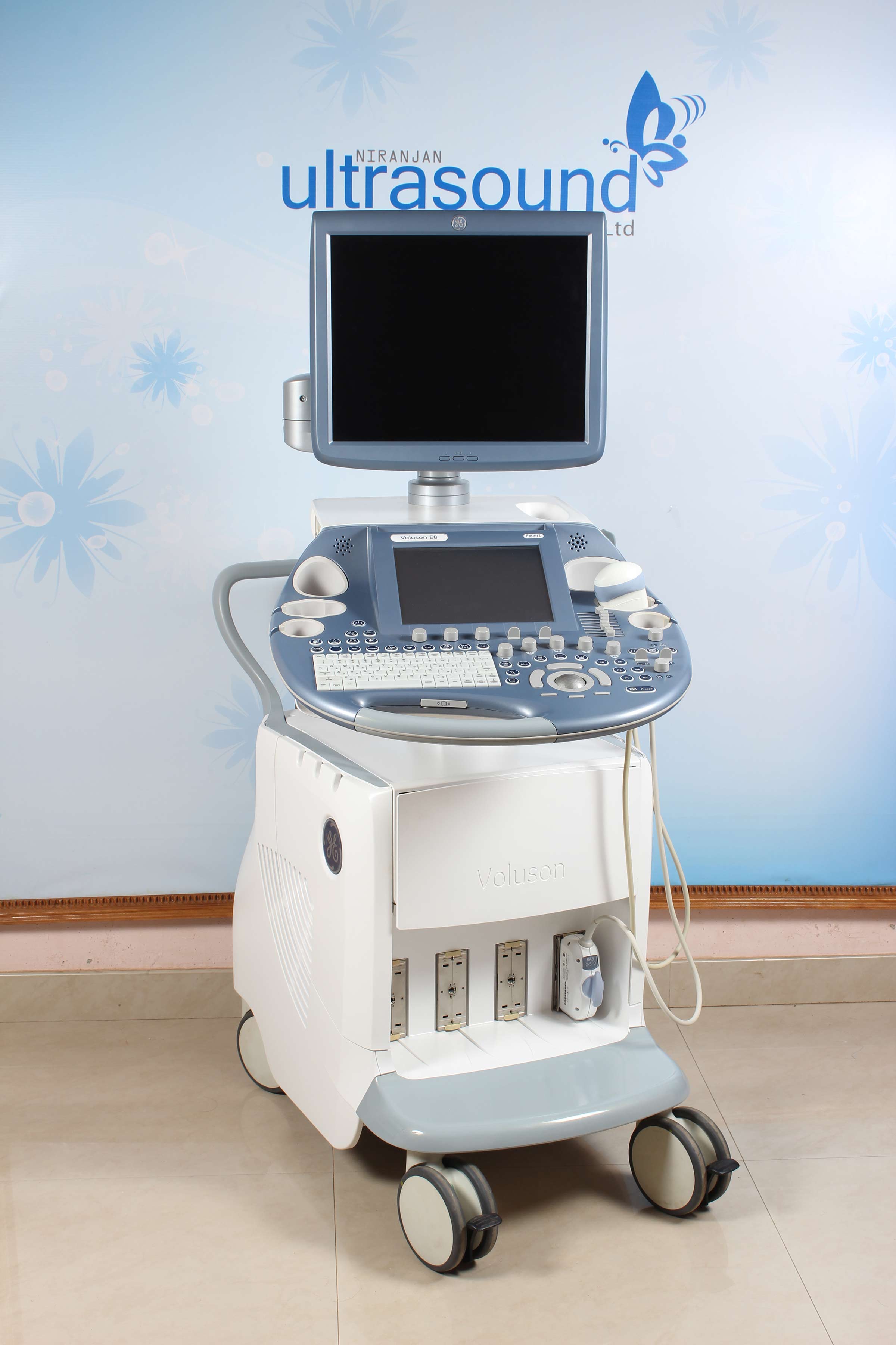
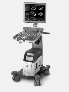
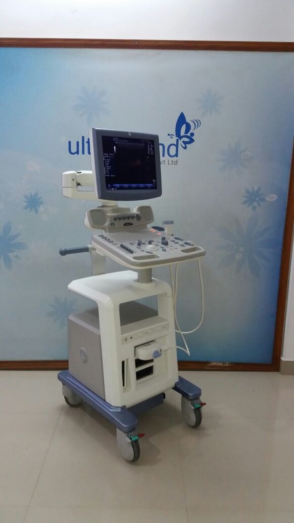
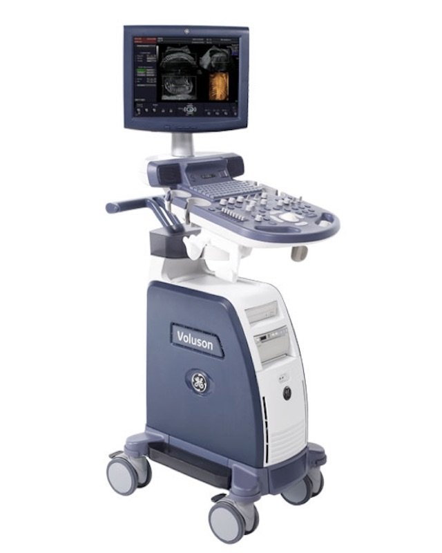
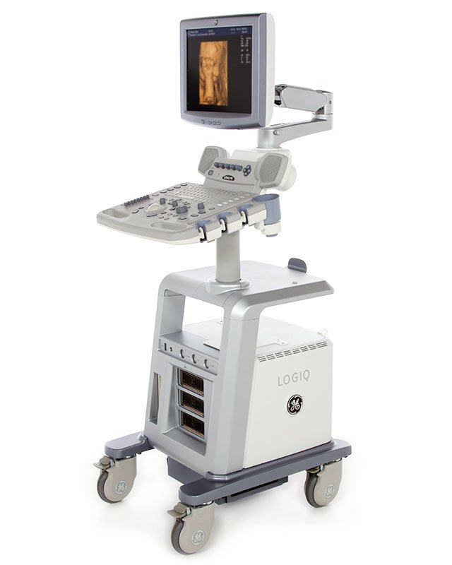
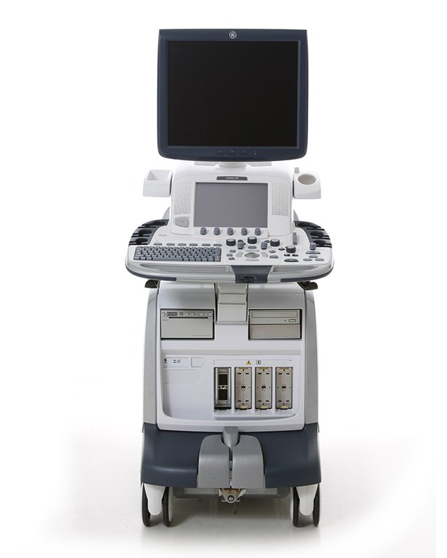
GE Voluson E8 BT 13
Year of Manufacture : February 2013
Software Version : BT 13.0.1
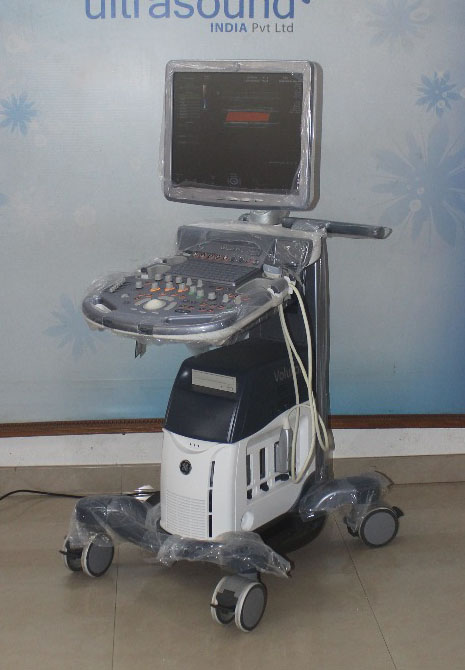
Philips IE33 F Cart
Year of Manufacture : 2010
Software Version : 5.0.0
GE Voluson E8 BT10
Year of Manufacture : 2011
Software Version : 10.0.0 BT10
GE Voluson E6 BT13.5
Year of Manufacture : 2014
Software Version : 14.0.2 BT13.5
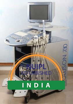
GE VOLUSON 730 PRO BT04
Year of Manufacture : November 2006
Software Version : 5.2.0
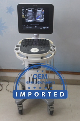
PHILIPS HD Clear Vue 550
Year of Manufacture : 2014
Software Version : 1.0.1
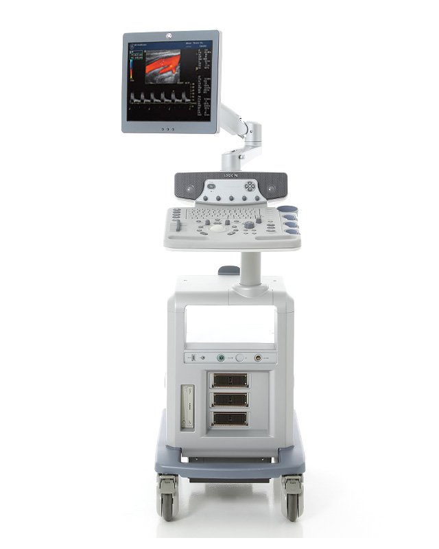
GE Logiq P6 PRO
Year of Manufacture : 2013
Software Version : 3.0.6
Ge Voluson S6
Year of Manufacture : Oct 2013
Software Version : A12.0.3
GE Voluson S8
Year of Manufacture : 2014
Software Version :
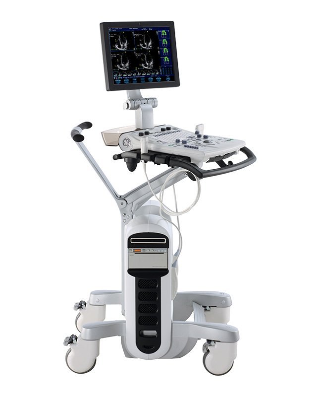
GE Logiq e
Year of Manufacture : July 2010
Software Version : R 5.0.1
Ge Voluson 730 Expert BT08
Year of Manufacture : Refurbished in 2017
Software Version : A. 5.4.6
GE Voluson P8
Year of Manufacture : Refurbished in 2017
Software Version : 13.0.0
Ge Voluson E8 – Radiance BT16
Year of Manufacture : Dec-2015
Software Version : 16.0.1 BT16
Philips IE33
Year of Manufacture :
Software Version : 5.0.0.318
GE VOLUSON 730 PRO
Year of Manufacture : April 2011
Software Version : 5.4.6
GE Voluson E8 BT13.5
Year of Manufacture : 2015
Software Version :
GE Voluson S8
Year of Manufacture : 2014
Software Version :
GE Logiq P5
Year of Manufacture : 2013
Software Version :
GE Voluson S8
Year of Manufacture : 2014
Software Version :
GE Voluson S8
Year of Manufacture : 2013
Software Version :
GE Voluson S8
Year of Manufacture : 2014
Software Version :
GE Voluson S8
Year of Manufacture : 2013
Software Version :
GE Voluson S6
Year of Manufacture : 2013
Software Version :
Ge Voluson E8 BT13
Year of Manufacture : 2013
Software Version :
GE Voluson E6 BT13.5
Year of Manufacture :
Software Version :
GE Voluson E6 BT13
Year of Manufacture : 2013
Software Version :
Ge Logiq P5
Year of Manufacture : 2012
Software Version :
Ge Logiq P5
Year of Manufacture : 2010
Software Version :
Ge Logiq P5
Year of Manufacture : 2010
Software Version :
GE Logiq P5
Year of Manufacture : 2009
Software Version :
Ge Logiq F8
Year of Manufacture : 2015
Software Version :
Ge Logiq F8
Year of Manufacture : 2015
Software Version :
Ge Logiq S8 XD Clear
Year of Manufacture : 2015
Software Version :
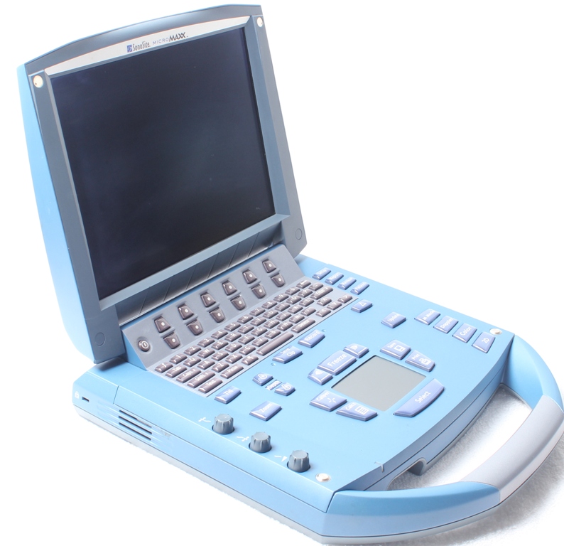
Sonosite MicroMaxx
Year of Manufacture : 2010-06
Software Version : 30.80.306.030
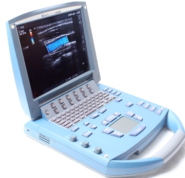
SONOSITE MICROMAXX
Year of Manufacture : 2011-08
Software Version : 30.80.306.024
SONOSITE MICROMAXX
Year of Manufacture : 2011 August
Software Version : 30.80.306.024
Voluson E6
Year of Manufacture : Oct-11
Software Version : 13.0.3 (BT13)
VOLUSON E6
Year of Manufacture : Feb-15
Software Version : 14.0.6 (BT13.5)
VOLUSON E8
Year of Manufacture : Apr-14
Software Version : 14.0.8 (BT13.5)
VOLUSON S8
Year of Manufacture : May-12
Software Version : 11.0.5
VOLUSON S6
Year of Manufacture : Jan-13
Software Version : 12.0.3
VOLUSON P8
Year of Manufacture : 2012
Software Version : 13.0.2
LOGIQ P5
Year of Manufacture : Oct-10
Software Version : 4.0.2
LOGIQ P6
Year of Manufacture : Dec-13
Software Version :
VIVID S5
Year of Manufacture : Oct-09
Software Version : 3.0.10
VIVID S6
Year of Manufacture : Apr-11
Software Version : 6.0.9
VIVID E9 XD Clear
Year of Manufacture : Jun-14
Software Version : 104.3.6
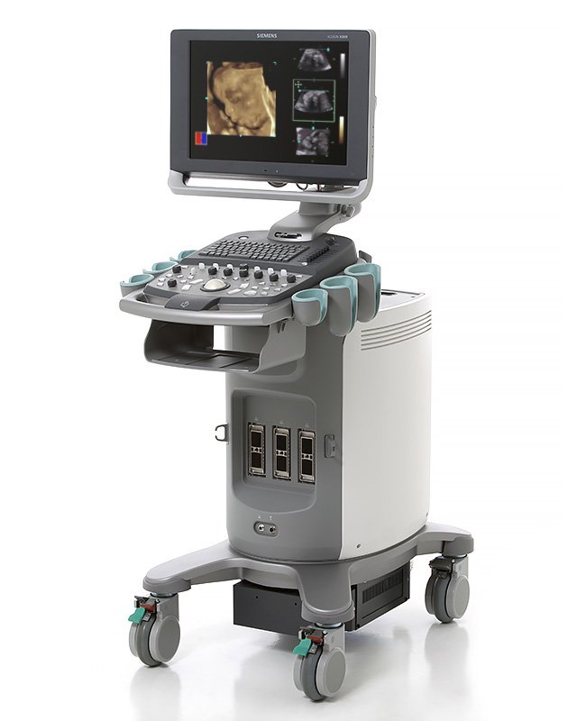
ACUSON X300 PE
Year of Manufacture : Nov-12
Software Version : 7.0.04
ACUSON X300 PE
Year of Manufacture : Oct-12
Software Version : 7.0.04
Niranjan and India Ultrasound team,
We are preparing to fly to Gujarat this morning. I would like to take this opportunity to say Thank you for an opportunity to a life time memory visiting India Ultrasound and South India.
Dear Sir wanted to thank you for the uncomplicated handling of the ultrasound - machine deal. I also appreciated the visit of the two very kind and friendly technicians who came here - to the "very remote" area, to do the necessary. The only little difficulty was, that your mails are put in the SPAM file - for whatever reason.
Mr.Niranjan
Team Sabisu thanks team IUS for the courtesy extended during our visit to your office for the event of CD training. It was wonderful and informative. Team Sabisu is highly motivated and ready to fire by all cylinders. Our spl. Thanks to Mr.Jithish for his warm guidance and reception.
Dear Sirs Received the probe today evening in excellent working condition & I must thank u for the same. The service which I received was excellent & prompt.
The equipment Logiq 5PRO arrive in Cape Verde.the equipmente arrived in very good conditions, and very good cosmetics .
Through you I want convey Our Heartiest Thanks and Regards to M/S India ultrasound India , For Repairing 3.5 Mega Hz Convex Probe of L&T ltrasound machine of Distt Hospital Agra ( UP),Free of cost.
Dear Niranjan, It was really nice to have seen your facility last Saturday. Your people did a great job of showing me around and I obtained all the vital information that I needed to know.
Have received machine. Still using it. No problems have faced yet. Overall satisfied.
Hello Sir,
It’s wonderful experience. Well organised, Highly Informative and very comfortable hospitality I have ever felt in Domestic sector.I wonder about your High Calibre Team Work and the Quality you all deliver not only in product, but in every aspect.As a whole the Leadership by Mr. NIRANJAN is awesome. In simple words, “I found the good seeds he sowed and hence, he reaps SUCCESS”.
Dear Niranjan,
Thank you very much for your reliable and honest business. As your faithful customer, I'm looking forward for further collaboration.
Dear Joshy
First of all I really want to thank you and all your team for all the efforts you guys have made to finally get the equipment where it belongs to.
Hi Niranjan how are you? we have received the systems and they look really good, thank you the plastic you put on the footrest and keyboard what is this. it is really nice to have.
Dear Ms. Binsy / Mr. Joshy & Mr. Niranjan
I’d like to thank you to send me prices, photos and videos. Also, I’d like to say that your job is extremely professional. I haven’t from others dealers this great job that you do, sending prices, pictures and now videos.
Dear Niranjan
Thank you for the courtesy extended to us during our visit to your place. You have a nice team there. the visit gave us a real good insight into the operation of the ultrasound machine and its features.
thanks and regards
Till now all ok, Satisfied with our company, Right now on a deal for logiq 100 pro with us.
Greetings prasanth,
The Logiq P5 has arrive in Montego Bay, Jamaica. I will go to the Sangster's Airport tomorrow to collect the Loqiq P5.
Hi, I arrived well and I am grateful for your kind reception and must say I enjoyed my visit to Kerala. My best wishes and please keep in touch.
Probe is still using. No complaints have gone through yet
Dear Maniji/ Joshiji/Niranjanji, Grateful thanks for sending your Engineer Mr.Navas P I will like to request that you kindly start a centre here.I can provide you all necessary infrastucture at almost NIL cost.I had expressed the same to Mr Navas
than u for your quick services. the probe images are good. small linear crack is there, but mr Bini raj told that it wont cause any trouble. pl send the invoice sos after the cheaque is cleared.pl also send the warrant letter. pl start ur repairing services in delhi also, it would be quite beneficial to northern region.
Dear Joshy, We got the machine successfully. Everything is fine, we really like it. We are very pleased to be working with you. We have one more request. We need a service code, and service manual. -- Best regards
Thank you, Mani. Everything worked. Pleasure doing business with you again.
Satsified with product, It was delivered in time frame.I have had 2 positive experiences with your company and will not hesitate to reccomend you to my colleages , trading internationally can prove difficult it is refreshing to find a company that does hold its values.Thank you
Machine is working good. Satisfied with the company, No problems hve faced yet
No compliants yet. Will surely contact for future deal
Machine and probes working absolutely fine, no issues.
I'm very much obliged to you. This trip for me was a very good experience, experience and familiarity with the kind , hospitable and Honest people. Your training was very good for me because i gained new experiences in the field of ultrasound. I wish you many years of happiness. I wish you many years of happiness.
Your company does tremendous work. The last three systems you sent us were perfect, which is consistent with every refurbished system I have bought from you over the years.
Hi Jameer, everything is done. Sarath had come on time n machine is fully functional. N thanks for ur kind support.
Dear Mani, The direction you gave me on installing a new battery successfully solved the problem I have been complaining about. You are really an asset to your company. How I wish we have someone who knows his ultrasound engineering like you somewhere near my locality. Perhaps the Logiq 200 would have been fixed easily.
Very prompt, courteous and efficient sales & service, I already bought 4 units, 2 for myself (GE Logiq e & P3) and 2 for my friends (one Logiq e and one P5). All of them are working well with minimal maintenance. I would recommend this company to any one with out hesitation .
Dear Joshy,
Today I have received the Logiq e and everything is in working order. Just wanted to say thanks for doing business. I am happy very with the purchase and will recommend you to my orthopaedic colleagues who are looking into purchasing an (non-portable) ultrasound too and happy to spread the word.


 +91 9847 069 684